
Lucía Manso-Ortega (1) y Jennifer Balade-González (2)
(1) Dept. de Lengua Vasca y Comunicación, Universidad del País Vasco, España
(2) Dept. de Psicología Cognitiva, Social y Organizacional, Universidad de La Laguna, España
(cc) Lucía Manso.
When a brain tumor grows near language areas in a multilingual patient, surgery goes beyond a mere medical procedure. Direct cortical electrostimulation mapping allows surgeons to build, in real time and with the patient awake, a unique functional map for each brain. Research shows that in multilingual brains, each language occupies partially distinct cortical areas, meaning that evaluating only one language during surgery may preserve one while irreversibly damaging another. Protecting a patient’s full linguistic identity requires detailed preoperative planning and multidisciplinary teams, because losing a language is not just losing words.
There are scientific questions that are born in a laboratory, and others that arise from the real needs of patients. This is one of the latter.
How does the human brain organize language in multilingual people remains one of the great open questions in neuroscience. Do they share the same circuits? Do they overlap? Do they separate completely? These are fascinating questions, but they take on an entirely different importance when the patient in front of you speaks three languages and has a tumor growing near the areas that support them.
For decades, neuroscience assumed that language was localized in well-defined areas: Broca’s area for producing it, Wernicke’s area for understanding it. Following this logic, the entire brain could be parceled into regions with clear borders. This picture is pedagogically useful, but incorrect. We now know that the brain is a complex system of interconnected networks (Sporns, 2013), whose organization varies from person to person. What is resectable tissue in one patient may be essential for speaking or reading in another.
Low-grade gliomas are a type of brain tumor characterized by slow growth, but capable of interfering with cognitive functions. Treatment typically includes surgery with a clear objective: to remove as much tumor tissue as possible while preserving the patient’s functionality. But how do we draw that line?
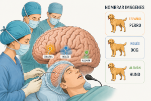
The most precise answer we have today is called direct cortical electrostimulation mapping (ESM; Ojemann, 1983). It is also one of the most demanding techniques, as it requires the patient to remain awake during part of the procedure. With the brain exposed, the surgeon applies brief, controlled, and reversible electrical currents to the cortical surface while the patient performs tasks: naming images or reading aloud, for example. If the stimulation of a specific area prevents the patient from completing the task, that is, if the function is interrupted, that area is considered essential. The moment the electrical current is removed, the function returns. In this way, a unique functional map is built for each patient, in real time, inside the operating room, and used to determine how much of the tumor can safely be removed. This approach has significantly extended the life expectancy of these patients.
For many years, standard practice was to evaluate only one language during surgery, under the assumption that this would be sufficient to protect language as a whole. The logic seemed reasonable, but as early as 1978, Ojemann and Whitaker described something that surprised the medical and scientific community: in bilingual patients, there were cortical zones specific to each language. Points at which stimulation interrupted one language while leaving the other completely intact.
We continue to observe this today (Martín-Fernández et al., 2022). The evidence tells us that languages in the bilingual brain are not completely separate, they share networks, particularly in temporal and parietal regions, but neither do they overlap entirely (Abutalebi & Green., 2016). The proportion of shared and language-specific zones varies enormously between individuals and appears to depend on factors such as the age of acquisition, proficiency or even the structural differences between the languages themselves.
The clinical implication is hard to ignore: if only one language is evaluated during surgery, there is a real risk of preserving one while irreversibly damaging another. After an apparently successful procedure, a patient might wake up unable to speak in one of their languages while the others remain intact. This selective loss is known as postoperative selective aphasia, and it illustrates just how much is at stake in preoperative planning.
Much of the work that makes intraoperative mapping possible begins long before the patient enters the operating room. Designing the cognitive battery, the tasks the patient will perform during stimulation, demands meticulous preparation. The tests must be brief, because time in the operating room is extremely limited; they must be balanced across languages and sensitive to different linguistic levels, such as phonological and semantic processing. In multilingual patients, the variables multiply, for example, which language is evaluated first.
Answering these questions requires multidisciplinary teams: surgeons, neurologists, neuropsychologists, speech therapists, linguists, and rehabilitation specialists, who plan together before the patient ever enters the operating room (Manso Ortega., 2022). For many people, losing a language is not just losing vocabulary, it can mean losing part of their identity.
Awake surgery is a highly personalized procedure that works with the neuroplasticity already taking place in the patient’s brain. The patient speaks, the team listens, and the surgery adapts in real time. In the end, the most important instrument in the operating room is the patient’s voice.
References
Abutalebi, J., & Green, D. W. (2016). Neuroimaging of language control in bilinguals: neural adaptation and reserve. Bilingualism: Language and Cognition, 19, 689–698.
Sporns O. (2013). Structure and function of complex brain networks. Dialogues in Clinical Neuroscience, 15, 247-62.
Ojemann, G. A., & Whitaker, H. A. (1978). The bilingual brain. Archives of Neurology, 35, 409–412.
Ojemann, G. A. (1983). Brain organization for language from the perspective of electrical stimulation mapping. Behavioral and Brain Sciences, 6, 189–206.
Manso-Ortega, L., et al. (2022). Highlighting the lack of neuropsychologists and speech therapists in healthcare services towards an accurate (pre- and postoperative) cognitive assessment in low-grade glioma patients. Psycho-oncology, 31, 1261–1263.
Martín-Fernández, J., et al. (2022). Intraoperative brain mapping in multilingual patients: What do we know and where are we going? Brain Sciences, 12, 560.
Manuscript received on May 13th, 2026.
Accepted on June 11th, 2026.
This is the English version of
Manso-Ortega, L., y Balade-González, J. (2026). ¿Cómo se representa el lenguaje en un cerebro multilingüe? Una respuesta desde el quirófano. Ciencia Cognitiva, 20:2, 49-51.
